21 November 2008

Rhinoplasty (croked nose ) -2

Pre- and pos-operative photography


Operative technique
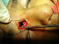
Ø The cartilaginous part of the hump is incised first, either under direct vision using an Aufricht retractor, or by palpation.
Ø Using the widest osteotome that can be inserted (14 mm or 16 mm in most patients) reduces the risk of an uneven resection of the hump. Rounded ends of the cutting edge reduce the risk of skin perforations.
Ø Whenever possible, the cartilaginous and bony part of the hump should be removed in one piece.
Ø The cut edges of the nasal bone are smoothed with a rasp, preferrably with a tungsten-carbide tip. By sliding the wet index finger over the dorsum, irregularities and insufficient resections may be palpated and corrected.
Ø The nasal bones are mobilised cranially by paramedian oblique osteotomies with a 2 mm or 3 mm osteotome being inserted through the nostril of the opposide side. The osteotome is driven in a latero-cranial direction towards a point that lies approximately 1 cm medial and rostral to the medial canthus.
Ø For the lateral osteotomy, the osteotome is placed on the piriform crest at the level of the insertion of the inferior turbinate by perforating the skin parallel to the piriform crest and then rotating the osteotome by 90°. The osteotome is driven towards the end of the paramedian oblique osteotomy, staying as far lateral as possible. In most patients the osteotome will cleave the bone ahead of the osteotome connecting the two osteotomy lines well before the tip of the osteotome reaches the end of the paramedian oblique osteotomy. At this moment the palpating finger will feel that the nasal bone “gives in” and the pitch of sound of the mallet striking the osteotome changes from high to low. In the great majority of patients, the nasal bone is now sufficiantly mobile with the remaining persiosteum acting as a hinge keeping the lateral aspect of the mobilised nasal bone from falling medially. The index finger and thumb palpate the position of the osteotome.
Ø Bruising and swelling of the eylids can be reduced by moderate pressure over the osteotomy and immediate placement of a cast or splint with digital pressure maintained.

Rhinoplasty 1 ( alar reduction)

Rhinoplasty, Alar Cartilage Resection


Use the least amount of infiltrative anesthesia (0.5% Xylocaine with 1:200,000 epinephrine) to prevent distortion of the nasal anatomy.
The tip cartilage can be approached by external approach - Bilateral marginal incisions connected by a transcolumellar incision

  • Removal of cephalic margin of the lateral crura
  • Suturing together of dome areas
  • Trimming of the caudal margin of medial crura
  • Insertion of a columellar strut
  • Larg excision of the alar to the vestibule around the alar facial junction to correct widely flaring alas. The scar should be kept approximately 1 mm on the alar side of the alar facial crease.


18 November 2008

Endoscopic Adeniodectomy

Operative technique

The Operative instruments include 70° and 30° rigid endoscope ,straight and up-biting angled forceps , adenoid curettage, angled suction diathermy, angled suction for maxillary sinus, Boyle–Davis mouth gag and plastic suction catheter .
The patient is placed in a supine position with the neck fully extended. After intubating the patient with an oro-endotracheal tube, is applied to keep the mouth open. The soft palate is palpated for the presence of a submucosal cleft. The posterior and lateral nasopharyngeal walls are palpated for pulsation. A plastic suction catheter is passed through each nostril and retrieved through the mouth , mild traction is needed to see the nasopharynx .
The surgeon stands on the head side of the patient and a 70° endoscope to examine the nasopharynx.


In some cases as in choanal adenoid and in the tubal tonsils , the surgeon stands on the right side of the patient .

A 30° rigid endoscope is used to evaluate the nasopharynx and Cotton pledgets with a mixture of 4% lidocaine and 1:100,000 epinephrine are placed in the nasal cavities to shrink the nasal mucosa. Then a transnasal adenoidectomy is done “Adenoid tissue in the nasopharynx was removed under direct visualization with straight or up-biting forceps”.

During the EA, the patient’s Eustachian tube orifices were visualized, and damage to this area was avoided. Once the patient’s adenoids had been removed, hemostasis was then obtained transorally and transnasally as indicated.

And in case of submucosal cleft palate, we perform an upper 1/2 ‘‘partial’’ adenoidectomy, combined transoral endoscopic adenoidectomy and transnasal endoscopic adenoidectomy was done.

Haemostasis was done by angled suction diathermy and the nasopharyngeal pack and revaluation of what was been removed and removal of the residual tissue is done on table, this can be repeated if needed.


FB in the oesophegous (oesphegoscopy)1


FB in the trachea (bronchoscopy)


Nasolabial cyst excision


frantoethmoidal mucocele

Preopretive CT
Operative finding
under endoscopic visualization using 2.7-mm 30 degrees telescopes. The middle turbinate is gently retracted medially to provide exposure of the bulla ethmoidalis alone.
Partial uncinectomy was performed to provide exposure of the anterior and medial walls of the bulla ethmoidalis, but this is later discontinued as access to the medial wall of the bulla ethmoidalis proved sufficient.
After completing an ethmoidectomy using the anterior-to-posterior, The medial wall of the bulla ethmoidalis was removed, including the posteriorly located natural ostium. The technique initially involved using a 90 degrees pediatric up-biting cup . than frontal recess become accessible and the frontal sinus easily opened. this lead to wide drainage of the mucus into the ethmoid cavity
Using a sickle knife , lamina papyracea fractured to provide restore of the eye in position

the extra bone formation due to the long stanging irrtation was removed to facilltate the movement of the eye.

However, exposure and drainage of this site is difficult, after derange and sending the material for gram staining and ap­propriate microbiologic studies, the cavity should be irrigated vigorously with saline, and the procedure is completed with the placement of derange tube to prevent the closure of the frontal sins and recollection again.
Postoperative picture