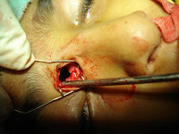

Operative finding


 under endoscopic visualization using 2.7-mm 30 degrees telescopes. The middle turbinate is gently retracted medially to provide exposure of the bulla ethmoidalis alone.
Partial uncinectomy was performed to provide exposure of the anterior and medial walls of the bulla ethmoidalis, but this is later discontinued as access to the medial wall of the bulla ethmoidalis proved sufficient.
 After completing an ethmoidectomy using the anterior-to-posterior, The medial wall of the bulla ethmoidalis was removed, including the posteriorly located natural ostium. The technique initially involved using a 90 degrees pediatric up-biting cup . than frontal recess become accessible and the frontal sinus easily opened. this lead to wide drainage of the mucus into the ethmoid cavity


 the extra bone formation due to the long stanging irrtation was removed to facilltate the movement of the eye.

 However, exposure and drainage of this site is difficult, after derange and sending the material for gram staining and appropriate microbiologic studies, the cavity should be irrigated vigorously with saline, and the procedure is completed with the placement of derange tube to prevent the closure of the frontal sins and recollection again.
 After completing an ethmoidectomy using the anterior-to-posterior, The medial wall of the bulla ethmoidalis was removed, including the posteriorly located natural ostium. The technique initially involved using a 90 degrees pediatric up-biting cup . than frontal recess become accessible and the frontal sinus easily opened. this lead to wide drainage of the mucus into the ethmoid cavity

Using a sickle knife , lamina papyracea fractured to provide restore of the eye in position

 the extra bone formation due to the long stanging irrtation was removed to facilltate the movement of the eye.
 However, exposure and drainage of this site is difficult, after derange and sending the material for gram staining and appropriate microbiologic studies, the cavity should be irrigated vigorously with saline, and the procedure is completed with the placement of derange tube to prevent the closure of the frontal sins and recollection again.
 Postoperative picture

No comments:
Post a Comment