28 December 2008

Fixation of fracture frontal bone with sinus obliteration

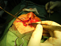
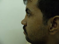
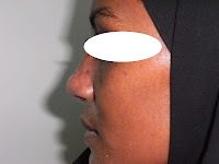
preoperative finding:
intraoprative data:

With displaced posterior table fractures, the risk of dural injury is unacceptably high.
In this case of frantal fracture , exploration for dural and repair was done.

To prevent late complications, these sinuse generally obliterated. The oblitrating material are muscle and fat from the thight.
With severe comminution of the posterior table, cranialization is the option.
A muscular flap can be used to separate the nasal and frontal cavities in patient as the cribriform plate was injured and thus augment the skull base and dural repair.
The anterior table can be reposioned and fixed by miniplate and scrow.
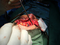
postoperative finding:


Treatment Options for fractures of the posterior table
Ø Nondisplaced without CSF leak
Observation
Ø Nondisplaced with CSF leak
Conservative management of CSF leak with progression to sinus exploration if no resolution in 4–7 days
Ø Displaced (>one table width)
Sinus exploration, repair of dura, obliteration or cranialization depending on involvement of the posterior table.
Ø Involvement of the nasofrontal outflow tract
Obliteration or~and cranialization

27 December 2008

Endoscopic management of subperiosteal orbital abscess

preoperative finding

operative techinque
After the administration of general anesthesia and intubation, cotton pads saturated with 0.05% oxymetazoline were applied to the anterior area of the nose.

Drainage of the SPOA was performed under endoscopic visualization using 2.7-mm and 4-mm 0 degrees and 30 degrees telescopes.
The middle turbinate is gently retracted medially to provide exposure of the bulla ethmoidalis alone. Partial uncinectomy was performed to provide exposure of the anterior and medial walls of the bulla ethmoidalis, but this is later discontinued as access to the medial wall of the bulla ethmoidalis proved sufficient. After completing an ethmoidectomy using the anterior-to-posterior or combined approach, careful homeostasis is obtained using packing saturated with a vasoconstrictive agent and bipolar cautery as needed


Because infection spreads from the ethmoid sinus, the lamina papyracea is often partially dehiscent in patients with a subperiosteal abscess. Using a bone curette, sufficient lamina papyracea should be removed to provide wide drainage of the abscess into the ethmoid cavity.


The periosteum can be incised in those rare instances where the abscess is within the adjacent orbit and drained into the ethmoid sinus. However, exposure and drainage of this site is difficult. After aspirating and sending the infectious material for gram staining and ap­propriate microbiologic studies, the abscess cavity should be irrigated vigorously with saline, and the procedure is completed without the placement of intrasinus packing and intranasal and intrasinus ointment.
posoperative finding


29 November 2008

Reconstruction of nasal skin loss(Double lobe flap)


Ø Bilobed flaps are used when the defect created by the primary flap (i.e., the banner flap) is too large to close primarily.
Ø The flaps are designed on a 45- to 90-degree axis to the primary defect and the flaps are elevated in the subcutaneous plane.
Ø Generally, the primary flap may be drawn somewhat smaller than the defect and is designed, as much as possible, to place the scars along minimal tension lines and within natural skin creases. Flaps designed at 45-degree angles minimize dog-ear formation.
Ø The resultant scars are complex and may be quite conspicuous.

28 November 2008

Rhinoplasty (augmentaion by Rib cartilage)

Rhinoplasty, Augmentation:

Intraoperative Details
Decongestion of the nasal mucosa with oxymetazoline, phenylephrine, allows adequate endonasal visualization. Injection of the nasal soft tissues with lidocaine and epinephrine allows adequate vasoconstriction and decreases intraoperative bleeding.
Augmentation performed via an open rhinoplasty approach. An open approach involves elevation of the skin and soft tissue envelope via a transcolumellar incision that is carried along the medial crura and the caudal aspect of the lower lateral cartilages. The entire cartilaginous skeleton of the lower and upper lateral cartilages should be exposed.
Dissection should proceed in a submuscular aponeurotic plane. Dissection superficial to this plane results in compromise of the vascular supply to the soft tissues and makes the dissection very difficult.
At the bony cartilaginous junction, the periosteum over the nasal bones is elevated and the dissection is carried in this plane up to the nasofrontal angle. The entire nasal skeleton should be adequately visualized.
Once the anatomy and the defects are adequately visualized harvesting of the rib cartilage was done. The rib is harvested through an incision along the chest wall to remove the cartilage portion of the rib, leaving behind the bony portion of the rib.
Grafts are fashioned and sculpted to the desired size and shape and are sutured in the desired location. The graft is secured in place with carefully placed, multiple 5-0 clear nylon.
Corrections are performed until the desired outcome is achieved. After augmentation is complete, the skin and soft tissue envelope are carefully red raped over the nasal skeleton and sutured in place with 6-0 nylon. An external nasal splint was performed.

pre- and post-operative finding:


27 November 2008

Rhinoplasty( crocked nase)

pre- and posoperative photography:


Intraoperative Details:

Correcting the deviated septum
A crooked nose is almost always associated with a crooked septum.
Deviating cartilage not included in the vital 1.5 cm dorsal caudal L strut can be resected without concern for loss of middle-vault support. Bony obstruction and deviations of the perpendicular plate of the ethmoid and vomer may be rongeured gently.
The caudal anterior septum is often displaced off the maxillary crest, which results in tip deviation. Corrective efforts include swinging the septum back onto its pedestal. This is performed by excising a triangular wedge of cartilage from the inferior septum at the posterior septal angle. The septal base is then stabilized to the maxillary crest periosteum with a strong, permanent suture.
recorrction through endonasal approach
In cases in which a twisted nose is accompanied by a lateral wall defect causing obstruction, treatment may be performed via the endonasal approach.
Although visualization is less than with the external rhinoplasty approach, combination of structural stenting and camouflaging may be adequate to achieve a straight dorsum. This approach allows for functional improvement without disrupting a well-supported tip.

Management of a crooked bony dorsum
A 2- or 3-mm straight osteotome is desirable; however, this small of an instrument has an associated learning curve. The 4-mm guarded osteotome is effective for lateral osteotomies and can be used with minimal trauma, especially when the guard is placed on the inner surface of the pyramid.
When indicated, medial osteotomies are performed before lateral osteotomies. This provides a safe back fracture and allows for a complete break. Incomplete breaks or green-stick fractures lead to bony irregularities and postoperative shifting of the bones. Medial osteotomies are performed with the aid of a 6-mm curved osteotome placed at the junction of the nasal bones and septum. The osteotome is directed 25° lateral from the midline. Medial osteotomies, however, are not always needed. If a large open roof exists following hump resection, bony walls may be mobilized fully with lateral osteotomies alone.
Lateral osteotomies are usually performed in a high-low-high direction. This path allows for protection of the nasal airway at the nasal base by initiating osteotomy at an elevated (high) position on the pyriform aperture. This leaves a triangle of bone at the base of the pyriform aperture (Webster triangle), avoiding compromise of the nasal airway width. The osteotome then travels in a low path along the mid dorsum to maximize narrowing and then back high to prevent over-narrowing of the nasal root. If the nasal bones do medialize, internal stabilization with splints may be necessary to prevent support without outward migration.